Tag: bacteria

  • Mosquitoes are Drawn to Your Skin Bacteria

    This month’s paper (from open access journal PLoS ONE) is yet again about the impact on our health of the bacteria which have decided to call our bodies home. But, instead of the bacteria living in our gut, this month is about the bacteria which live on our skin.

    It’s been known that the bacteria that live on our skin help give us our particular odors. So, the researchers wondered if the mosquitos responsible for passing malaria (Anopheles) were more or less drawn to different individuals based on the scent that our skin-borne bacteria impart upon us (also, for the record, before you freak out about bacteria on your skin, remember that like the bacteria in your gut, the bacteria on your skin are natural and play a key role in maintaining the health of your skin).

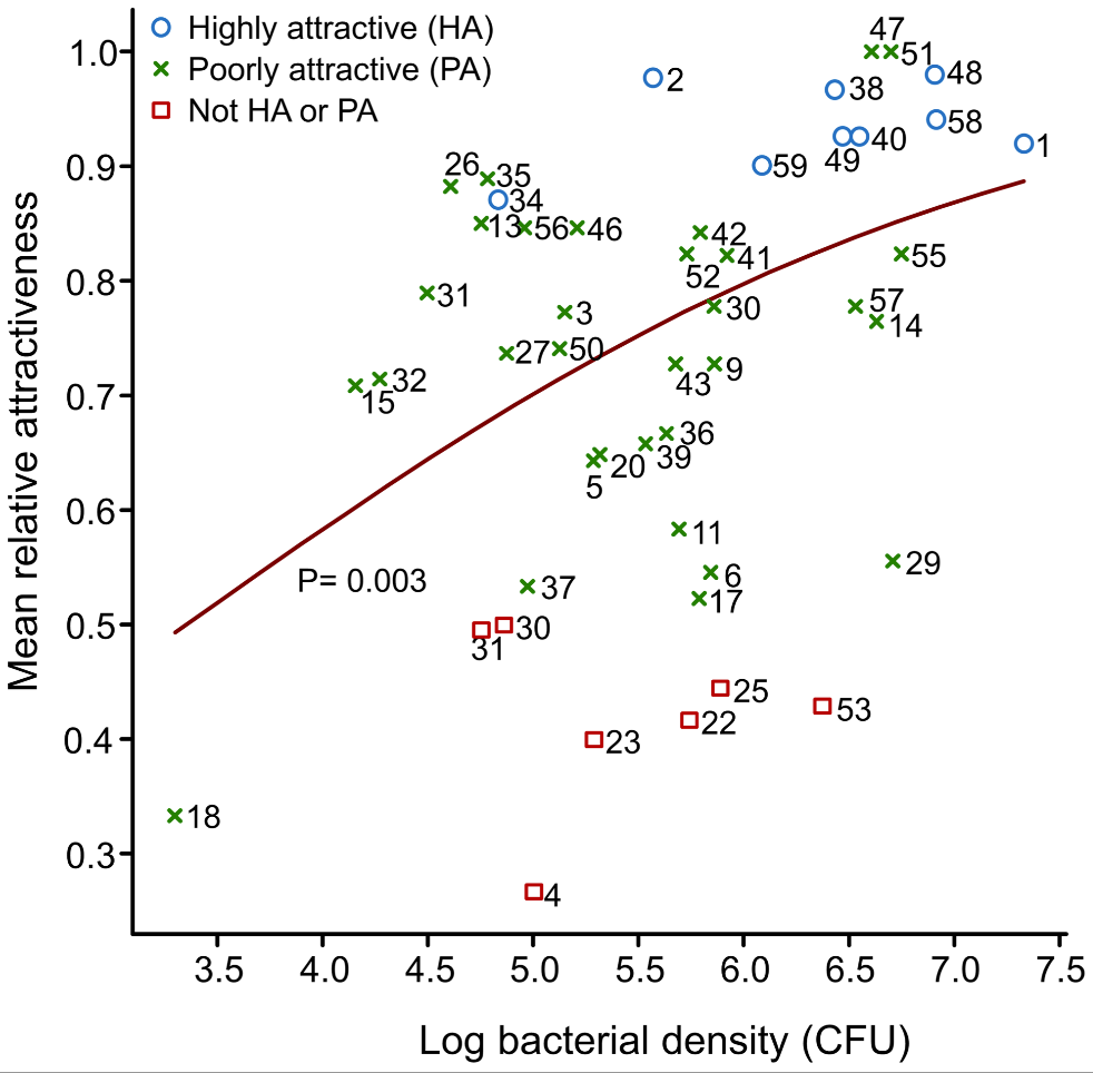
    Looking at 48 individuals, they noticed a huge variation in terms of attractiveness to Anopheles mosquitos (measured by seeing how much mosquitos prefer to fly towards a chamber with a particular individual’s skin extract versus a control) which they were able to trace to two things. The first is the amount of bacteria on your skin. As shown in Figure 2 below, is that the more bacteria that you have on your skin (the higher your “log bacterial density”), the more attractive you seem to be to mosquitos (the higher your mean relative attractiveness).

    Source: Figure 2, Verhulst et al

    The second thing they noticed was that the type of bacteria also seemed to be correlated with attractiveness to mosquitos. Using DNA sequencing technology, they were able to get a mini-census of what sort of bacteria were present on the skins of the different patients. Sadly, they didn’t show any pretty figures for the analysis they conducted on two common types of bacteria (Staphylococcus and Pseudomonas), but, to quote from the paper:

    The abundance of Staphylococcus spp. was 2.62 times higher in the HA [Highly Attractive to mosquitoes] group than in the PA [Poorly Attractive to mosquitoes] group and the abundance of Pseudomonas spp. 3.11 times higher in the PA group than in the HA group.

    Using further genetic analyses, they were also able to show a number of other types of bacteria that were correlated with one or the other.

    So, what did I think? While I think there’s a lot of interesting data here, I think the story could’ve been tighter. First and foremost, for obvious reasons, correlation does not mean causation. This was not a true controlled experiment – we don’t know for a fact if more/specific types of bacteria cause mosquitos to be drawn to them or if there’s something else that explains both the amount/type of bacteria and the attractiveness of an individual’s skin scent to a mosquito. Secondly, Figure 2 leaves much to be desired in terms of establishing a strong trendline. Yes, if I  squint (and ignore their very leading trendline) I can see a positive correlation – but truth be told, the scatterplot looks like a giant mess, especially if you include the red squares that go with “Not HA or PA”. For a future study, I think it’d be great if they could get around this to show stronger causation with direct experimentation (i.e. extracting the odorants from Staphylococcus and/or Pseudomonas and adding them to a “clean” skin sample, etc)

    With that said, I have to applaud the researchers for tackling a fascinating topic by taking a very different angle. Coverage of malaria is usually focused on how to directly kill or impede the parasite (Plasmodium falciparums). This is the first treatment of the “ecology” of malaria – specifically the ecology of the bacteria on your skin! While the authors don’t promise a “cure for malaria”, you can tell they are excited about what they’ve found and the potential to find ways other than killing parasites/mosquitos to help deal with malaria, and I look forward to seeing the other ways that our skin bacteria impact our lives.

    Paper: Verhulst et al. “Composition of Human Skin Microbiota Affects Attractiveness to Malaria Mosquitoes.” PLoS ONE 6(12). 17 Nov 2011. doi:10.1371/journal.pone.0028991

    Check out my other academic paper walkthroughs/summaries

  • Fat Flora

    Source: Healthy Soul

    November’s paper was published in Nature in 2006, and covers a topic I’ve become increasingly interested in: the impact of the bacteria that have colonized our bodies on our health (something I’ve blogged about here and here).

    The idea that our bodies are, in some ways, more bacteria than human (there are 10x more gut bacteria – or flora — than human cells on our bodies) and that those bacteria can play a key role on our health is not only mind-blowing, it opens up another potential area for medical/life sciences research and future medicines/treatments.

    In the paper, a genetics team from Washington University in St. Louis explored a very basic question: are the gut bacteria from obese individuals different from those from non-obese individuals? To study the question, they performed two types of analyses on a set of mice with a genetic defect leading to an inability of the mice to “feel full” (and hence likely to become obese) and genetically similar mice lacking that defect (the s0-called “wild type” control).

    The first was a series of genetic experiments comparing the bacteria found within the gut of obese mice with those from the gut of “wild-type” mice (this sort of comparison is something the field calls metagenomics). In doing so, the researchers noticed a number of key differences in the “genetic fingerprint” of the two sets of gut bacteria, especially in the genes involved in metabolism.

    Source: Figure 3, Turnbaugh et al.

    But, what did that mean to the overall health of the animal? To answer that question, the researchers did a number of experiments, two of which I will talk about below. First, they did a very simple chemical analysis (see figure 3b to the left) comparing the “leftover energy” in the waste (aka poop) of the obese mice to the waste of wild-type mice (and, yes, all of this was controlled for the amount of waste/poop). Lo and behold, the obese mice (the white bar) seemed to have gut bacteria which were significantly better at pulling calories out of the food, leaving less “leftover energy”.

    Source: Figure 3, Turnbaugh et al.

    While an interesting result, especially when thinking about some of the causes and effects of obesity, a skeptic might look at that data and say that its inconclusive about the role of gut bacteria in obesity – after all, obese mice could have all sorts of other changes which make them more efficient at pulling energy out of food. To address that, the researchers did a very elegant experiment involving fecal transplant: that’s right, colonize one mouse with the bacteria from another mouse (by transferring poop). The figure to the right (figure 3c) shows the results of the experiment. After two weeks, despite starting out at about the same weight and eating similar amounts of the same food, wild type mice that received bacteria from other wild type mice showed an increase in body fat of about 27%, whereas the wild type mice that received bacteria from the obese mice showed an increase of about 47%! Clearly, gut bacteria in obese mice are playing a key role in calorie uptake!

    In terms of areas of improvement, my main complaint about this study is just that it doesn’t go far enough. The paper never gets too deep on what exactly were the bacteria in each sample and we didn’t really get a sense of the real variation: how much do bacteria vary from mouse to mouse? Is it the completely different bacteria? Is it the same bacteria but different numbers? Is it the same bacteria but they’re each functioning differently? Do two obese mice have the same bacteria? What about a mouse that isn’t quite obese but not quite wild-type either? Furthermore, the paper doesn’t show us what happens if an obese mouse has its bacteria replaced with the bacteria from a wild-type mouse. These are all interesting questions that would really help researchers and doctors understand what is happening.

    But, despite all of that, this was a very interesting finding and has major implications for doctors and researchers in thinking about how our complicated flora impact and are impacted by our health.

    Paper: Turnbaugh et al., “An obesity-associated gut microbiome with increased capacity for energy harvest.” Nature (444). 21/28 Dec 2006. doi:10.1038/nature05414

    Check out my other academic paper walkthroughs/summaries

  • Collateral Damage by Mitochondria

    This month, I read a paper (HT: my ex-college roommate Eric) by a group from Beth Israel about systemic inflammatory response syndrome (SIRS) following serious injury. SIRS, which is more commonly understood/found as sepsis, happens when the entire body is on high “immune alert.” In the case of sepsis, this is usually due to an infection of some sort. While an immune response may be needed to control an internal infection, SIRS is dangerous because the immune system can cause a great deal of collateral damage, resulting in potentially organ failure and death.

    Whereas an infection has a clear link to sepsis, the logic for why injury would cause a similar immune response was less clear. In fact, for years, the best hypothesis from the medical community was that injury would somehow cause the bacteria which naturally live in your gut to appear where they’re not supposed to be. But this explanation was not especially convincing, especially in light of injuries like burns which could still lead to SIRS but which didn’t seem to directly affect gut bacteria.

    Source: The Brain From Top to Bottom

    Zhang et al, instead of assuming that some type of  endogenous bacteria was being released following injury, came up with an interesting hypothesis: it’s not bacteria which is triggering SIRS, but mitochondria. A first year cell biology student will be able to tell you that mitochondria are the parts of eukaryotic cells (sophisticated cells with nuclei) which are responsible for keeping the cell supplied with energy. A long-standing theory in the life science community (pictured above) is that mitochondria, billions of years ago, were originally bacteria which other, larger bacteria swallowed whole. Over countless rounds of evolution, these smaller bacteria became symbiotic with their “neighbor” and eventually adapted to servicing the larger cell’s energy needs. Despite this evolution, mitochondria have not lost all of their (theorized) bacterial ancestry, and in fact still retain bacteria-like DNA and structures. Zhang et al’s guess was that serious injuries could expose a mitochondria’s hidden bacterial nature to the immune system, and cause the body to trigger SIRS as a response.

    Interesting idea, but how do you prove it? The researchers were able to show that 15 major trauma patients with no open wounds or injuries to the gut had thousands of times more mitochondrial DNA  in their bloodstream than non-trauma victims. The researchers were then able to show that this mitochondrial DNA was capable of activating polymorphonuclear neutrophils, some of the body’s key “soldier” cells responsible for causing SIRS.

    Source: Figure 3, Zhang et al.

    The figure above shows the result of an experiments illustrating this effect looking at the levels of a protein called p38 MAPK which gets chemically modified into “p-p38” when neutrophils are activated. As you can see in the p-p38 row, adding more mitochondrial DNA (mtDNA, “-” columns) to a sample of neutrophils increases levels of p-p38 (bigger, darker splotch), but adding special DNA which blocks the neutrophil’s mtDNA “detectors” (ODN, “+” columns) seems to lower it again. Comparing this with the control p38 row right underneath shows that the increase in p-p38 is likely due to neutrophil activation from the cells detecting mitochondrial DNA, and not just because the sample had more neutrophils/more p38 (as the splotches in the second row are all roughly the same).

    Cool, but does this mean that mitochondrial DNA actually causes a strong immune response outside of a test tube environment? To test this, the researchers injected mitochondrial DNA into rats and ran a full set of screens on them. While the paper showed numerous charts pointing out how the injected rats had strong immune response across multiple organs, the most striking are the pictures below which show a cross-section of a rat’s lungs comparing rats injected with a buffer solution (panel a, “Sham”) and rats injected with mitochondrial DNA (panel b, MTD). The cross-sections are stained with hematoxylin and eosin which highlight the presence of cells. The darker and “thicker” color on the right shows that there are many more cells in the lungs of rats injected with mitochondrial DNA – most likely from neutrophils and other “soldier cells” which have rushed in looking for bacteria to fight.

    Source: Figure 4, Zhang et al.

    Amazing isn’t it? Not only did they provide part of the solution to the puzzle of injury-mediated SIRS (what they used to call “sterile SIRS”), but lent some support to the endosymbiont hypothesis!

    Paper: Zhang, Qin et al. “Circulating Mitochondrial DAMPs Cause Inflammatory Responses to Injury.” Nature 464, 104-108 (4 March 2010) – doi:10.1038/nature08780

    Check out my other academic paper walkthroughs/summaries